43 picture of compound microscope with labels
Microscope Parts and Functions Microscope Parts and Functions With Labeled Diagram and Functions How does a Compound Microscope Work?. Before exploring microscope parts and functions, you should probably understand that the compound light microscope is more complicated than just a microscope with more than one lens.. First, the purpose of a microscope is to magnify a small object or to magnify the fine details of a larger ... Binocular Microscope Anatomy - Parts and Functions with a Labeled ... Now, I will describe all these non-optical parts of the light compound microscope with the labeled diagrams. The body tube of the microscope. The body tube is the solid support for the optical and mechanical parts of the microscope. There are two basic types of stand in the body tube of a light compound microscope - upright stand and inverted ...
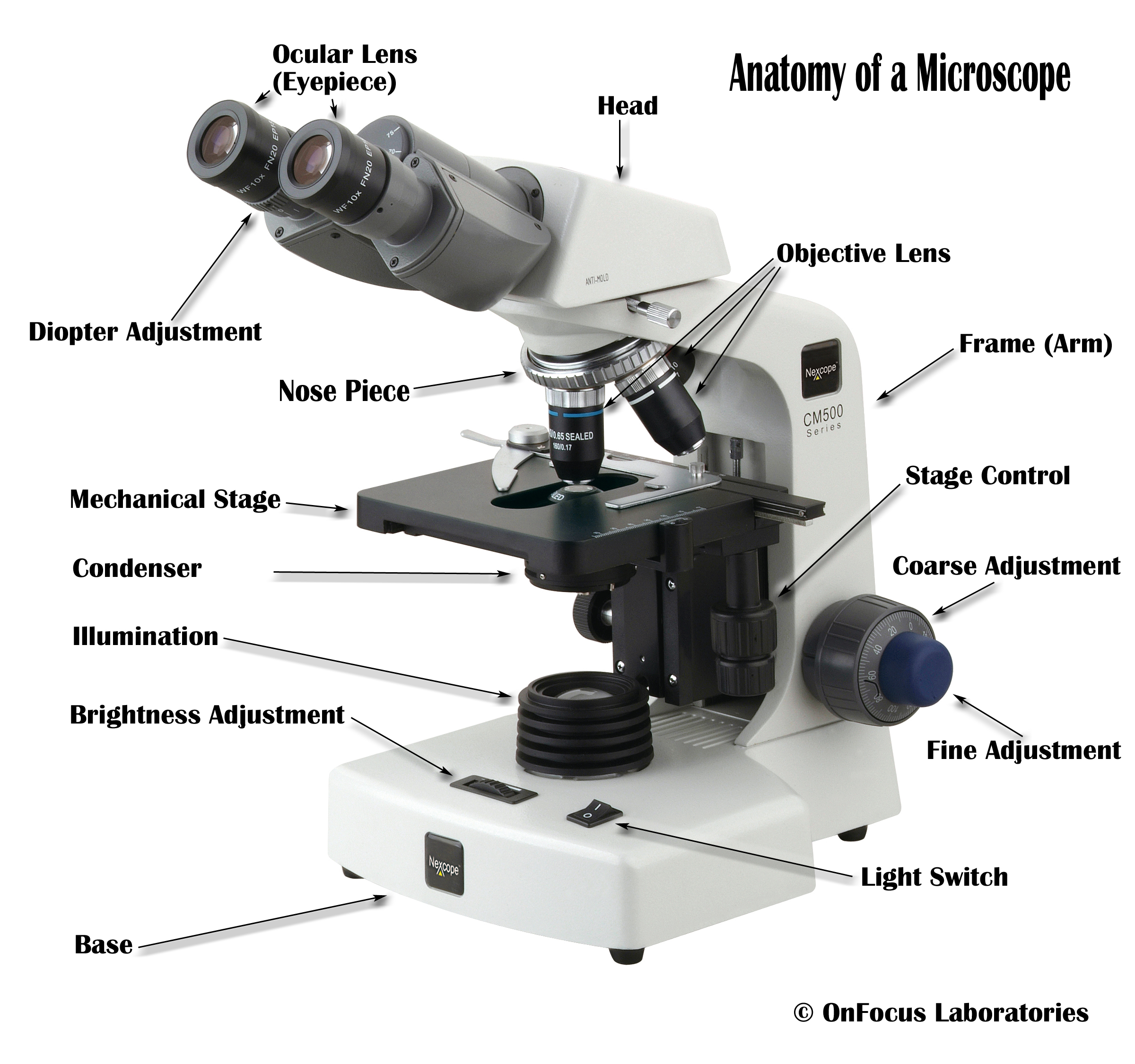
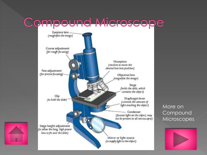
Compound Microscope - Diagram (Parts labelled), Principle and Uses See: Labeled Diagram showing differences between compound and simple microscope parts Structural Components The three structural components include 1. Head This is the upper part of the microscope that houses the optical parts 2. Arm This part connects the head with the base and provides stability to the microscope.

Picture of compound microscope with labels
Compound Microscope Labeled Diagram - Quizlet QUESTION. The total magnification of a specimen being viewed with a 10X ocular lens and a 40X objective lens is. 15 answers. QUESTION. a mosquito beats its wings up and down 600 times per second, which you hear as a very annoying 600 Hz sound. if the air outside is 20 C, how far would a sound wave travel between wing beats. 2 answers. Microscope Types (with labeled diagrams) and Functions A compound microscope: Is used to view samples that are not visible to the naked eye Uses two types of lenses - Objective and ocular lenses Has a higher level of magnification - Typically up to 2000x Is used in hospitals and forensic labs by scientists, biologists and researchers to study micro organisms Compound microscope labeled diagram Parts of a Compound Microscope - Labeled (with diagrams) A compound microscope is known as a high-power microscope that enables you to achieve a high level of magnification. Smaller specimens can be thoroughly viewed using a compound microscope. Let us take a look at the different parts of a compound microscope and understand each key component.
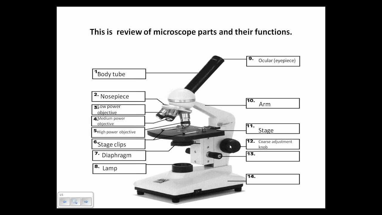
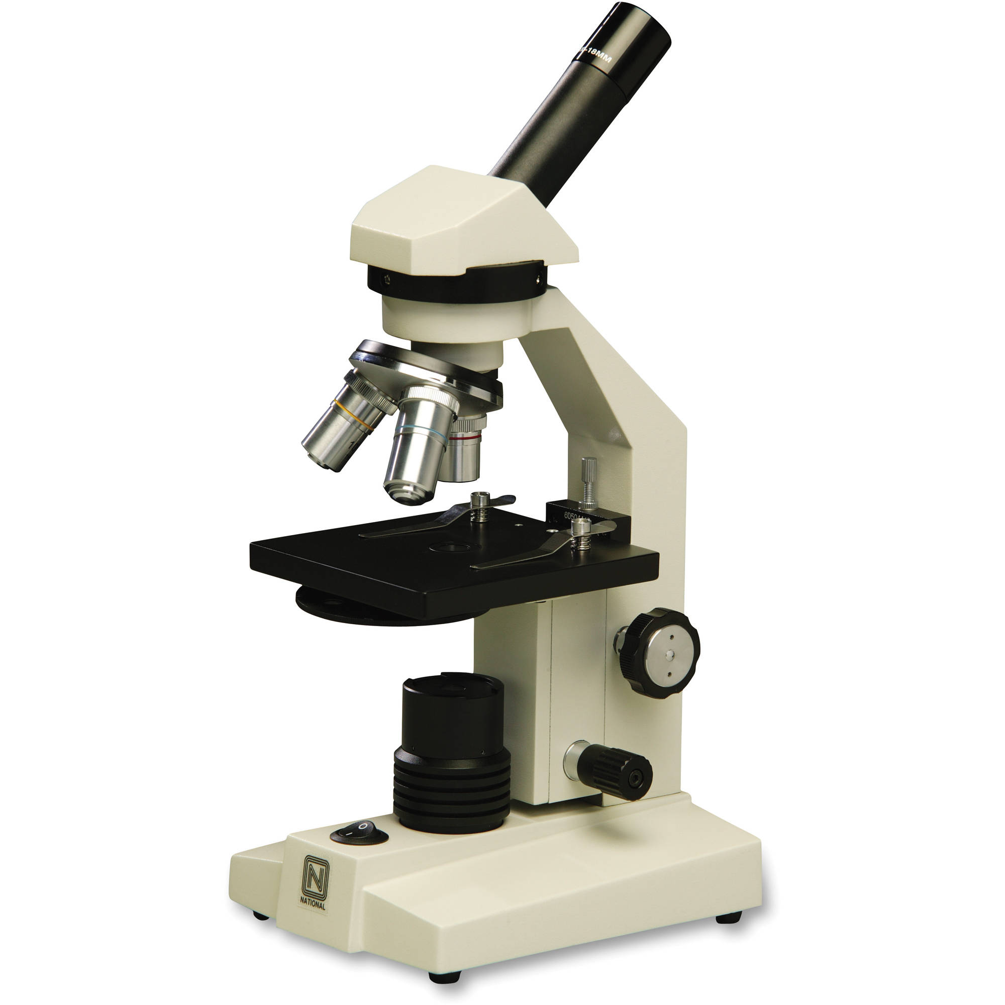
Picture of compound microscope with labels. Compound Microscope Parts Made Easy A compound microscope employs a system of lenses and light to magnify the specimen. ... There are usually 3 or 4 objective lenses per microscope (the picture above has 3), and they usually have powers of 4x, 10x, 40x, and 100x respectively (one power per lens). ... Not labeled in the above diagram but you can see it as a lightly glowing circle ... Labeling the Parts of the Microscope | Microscope activity, Science ... Funny Cartoons Description Worksheet identifying the parts of the compound light microscope. Answer key: 1. Body tube 2. Revolving nosepiece 3. Low power objective 4. Medium power objective 5. High power objective 6. Stage clips 7. Diaphragm 8. Light source 9. Eyepiece 10. Arm 11. Stage 12. Coarse adjustment knob 13. Fine adjustment knob 14. Base Compound Microscope: Definition, Diagram, Parts, Uses, Working ... - BYJUS A microscope with a high resolution and uses two sets of lenses providing a 2-dimensional image of the sample. The term compound refers to the usage of more than one lens in the microscope. Also, the compound microscope is one of the types of optical microscopes. The other type of optical microscope is a simple microscope. Compound Microscope Parts, Functions, and Labeled Diagram Compound Microscope Definitions for Labels. Eyepiece (ocular lens) with or without Pointer: The part that is looked through at the top of the compound microscope. Eyepieces typically have a magnification between 5x & 30x. Monocular or Binocular Head: Structural support that holds & connects the eyepieces to the objective lenses.
Compound Microscope- Definition, Labeled Diagram, Principle, Parts, Uses In order to ascertain the total magnification when viewing an image with a compound light microscope, take the power of the objective lens which is at 4x, 10x or 40x and multiply it by the power of the eyepiece which is typically 10x. Therefore, a 10x eyepiece used with a 40X objective lens will produce a magnification of 400X. Microscope Drawing And Label - Painting Valley Are you looking for the best images of Microscope Drawing And Label? Here you are! We collected 33+ Microscope Drawing And Label paintings in our online museum of paintings - PaintingValley.com. ... Compound Microscope ... 413x424 6 0. Like JPG. Microscope - Microsc... 236x262 4 1. Like JPG. Label Microscope Dia... 459x457 4 0. Like JPG ... Compound Microscope Stock Photos and Images - Alamy Find the perfect compound microscope stock photo. Huge collection, amazing choice, 100+ million high quality, affordable RF and RM images. ... Compound Microscope Stock Photos and Images (1,984) compound microscope isolated. Related searches: ... Method of illuminating compound microscope with gas lamp. Labels: C, ... Microscope picture label Flashcards | Quizlet Start studying Microscope picture label. Learn vocabulary, terms, and more with flashcards, games, and other study tools.
Compound microscope - their parts and function - Microscopy4kids Compound microscopes have more than one lens to generate high magnification images of flat, thin specimens. 2. Eyepiece (10x) and Objective lenses (4x, 10x, 40x, 100x) are two major optical parts of a microscope. 3. Total magnification power is calculated by multiplying the magnification of the eyepiece and objective lens. 4. Labelled Diagram of Compound Microscope - Biology Discussion The below mentioned article provides a labelled diagram of compound microscope. Part # 1. The Stand: The stand is made up of a heavy foot which carries a curved inclinable limb or arm bearing the body tube. The foot is generally horse shoe-shaped structure (Fig. 2) which rests on table top or any other surface on which the microscope in kept. A Study of the Microscope and its Functions With a Labeled Diagram To better understand the structure and function of a microscope, we need to take a look at the labeled microscope diagrams of the compound and electron microscope. These diagrams clearly explain the functioning of the microscopes along with their respective parts. ... The camera present within the microscope captures images to reveal the finer ... Labeling the Parts of the Microscope | Microscope World Resources Labeling the Parts of the Microscope This activity has been designed for use in homes and schools. Each microscope layout (both blank and the version with answers) are available as PDF downloads. You can view a more in-depth review of each part of the microscope here. Download the Label the Parts of the Microscope PDF printable version here.
labels of a compound microscope compound microscope labels 2324295 - Top Label Maker
Parts of the Microscope with Labeling (also Free Printouts) Microscopes are specially created to magnify the image of the subject being studied. This exercise is created to be used in homes and schools. the microscope layout, including the blank and answered versions are available as pdf downloads. Click to Download : Label the Parts of the Microscope (A4) PDF print version.

Microscopy
Diagram of a Compound Microscope - Biology Discussion 1. It is noted first that which objective lens is in use on the microscope. 2. Stage micrometer is positioned in such a way that it is in the field of view. 3. The eyepiece is rotated so that the two scales, the eyepiece or ocular scale and the stage micrometer scale, are parallel. 4.
23 Label And Color The Parts Of Both Microscopes - Labels 2021
Compound microscope Images, Stock Photos & Vectors - Shutterstock 3,117 compound microscope stock photos, vectors, and illustrations are available royalty-free. See compound microscope stock video clips Image type Orientation Sort by Popular Science College and University Biology Insects and Spiders Jobs/Professions microscope laboratory compound eye optical microscope scientist Next of 32

Compound Microscope, कंपाउंड माइक्रोस्कोप in Sector 24 , Uniteck Scientific & Electronic ...
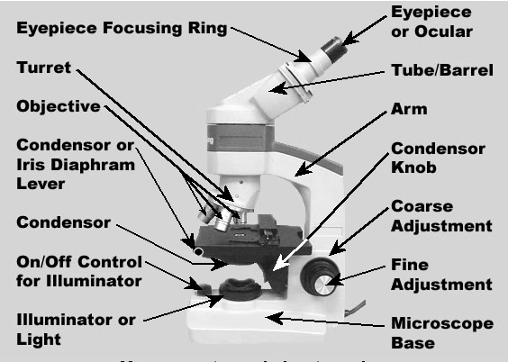
Parts of a microscope with functions and labeled diagram Q. Differentiate between a condenser and an Abbe condenser. Ans. Condensers are lenses that are used to collect and focus light from the illuminator into the specimen. They are found under the stage next to the diaphragm of the microscope. They play a major role in ensuring clear sharp images are produced with a high magnification of 400X and above.
Microscope Diagram to Print
Compound Microscope Parts - Labeled Diagram and their Functions - Rs ... Basically, compound microscopes generate magnified images through an aligned pair of the objective lens and the ocular lens. In contrast, "simple microscopes" have only one convex lens and function more like glass magnifiers. [In this figure] Two "antique" microscopes played significant roles in the history of biology.
1.Fully label the compound microscope below.
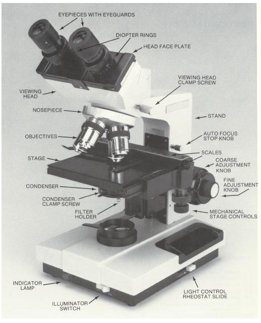
Compound Microscope - Types, Parts, Diagram, Functions and Uses Eyepiece/ocular lens - It is the part of the microscope that is looked through at the top. It comes with a magnification ranging between 5x and 30x. Image 3: The head connects the eyepiece to the objective lens. Picture Source: microscope.com. Head (monocular/binocular) - It is the structural support of the microscope.

Microscope Lab: The Compound Microscope by Amy Brown Science | TpT
26+ Picture Of A Microscope With Label PNG 26+ Picture Of A Microscope With Label PNG. Microscopes are specially created to magnify the image of the subject being studied. Students label the parts of the microscope in this photo of a basic laboratory light microscope. Microscope Drawing And Label at GetDrawings | Free download from getdrawings.com I searched for this on bing.com/images.

Microscope World Blog: Tonsils under the Microscope
Compound Microscope with labels Stock Vector | Adobe Stock Compound Microscope with labels Stock Vector | Adobe Stock. Get 10 free Adobe Stock images.

Anatomy and Physiology I Coursework: Microscope A+P
Parts of a Compound Microscope and Their Functions Compound microscope mechanical parts (Microscope Diagram: 2) include base or foot, pillar, arm, inclination joint, stage, clips, diaphragm, body tube, nose piece, coarse adjustment knob and fine adjustment knob. Base: It's the horseshoe-shaped base structure of microscope. All of the other components of the compound microscope are supported ...
Microscope Review.wmv - YouTube
A Study of the Microscope and its Functions With a Labeled Diagram May 21, 2019 - To better understand the structure and function of a microscope, we need to take a look at the labeled microscope diagrams of the compound and electron microscope. These diagrams clearly explain the functioning of the microscopes along with their respective parts.
Compound Microscope
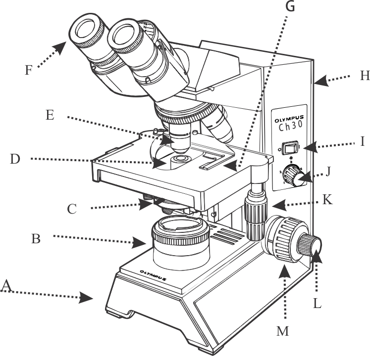
Label the microscope — Science Learning Hub All microscopes share features in common. In this interactive, you can label the different parts of a microscope. Use this with the Microscope parts activity to help students identify and label the main parts of a microscope and then describe their functions. Drag and drop the text labels onto the microscope diagram.

Anatomy and Physiology I Coursework: Microscope A+P
Parts of a Compound Microscope - Labeled (with diagrams) A compound microscope is known as a high-power microscope that enables you to achieve a high level of magnification. Smaller specimens can be thoroughly viewed using a compound microscope. Let us take a look at the different parts of a compound microscope and understand each key component.
33 Label Of Compound Microscope - Labels Database 2020
Microscope Types (with labeled diagrams) and Functions A compound microscope: Is used to view samples that are not visible to the naked eye Uses two types of lenses - Objective and ocular lenses Has a higher level of magnification - Typically up to 2000x Is used in hospitals and forensic labs by scientists, biologists and researchers to study micro organisms Compound microscope labeled diagram
PPT - Types of Microscopes PowerPoint Presentation - ID:1588009
Compound Microscope Labeled Diagram - Quizlet QUESTION. The total magnification of a specimen being viewed with a 10X ocular lens and a 40X objective lens is. 15 answers. QUESTION. a mosquito beats its wings up and down 600 times per second, which you hear as a very annoying 600 Hz sound. if the air outside is 20 C, how far would a sound wave travel between wing beats. 2 answers.
Microscope Lab

BIOLOGY BASICS - "Compound Microscope: Basic Use" - YouTube
Post a Comment for "43 picture of compound microscope with labels"